
Serological endpoints: Agglutination - Stage 2
Agglutination is the key end-point that we use in the vast majority of TML testing.
Agglutination occurs in two stages:
Stage 2 - Lattice formation: Once a red cell is coated with antibodies, the binding of the antibodies to multiple red cells causes visible red cell clumping to occur. The formation of a lattice requires the ability to overcome the natural repulsion between two neighbouring red cells. This electronic potential (or zeta potential) is due to the negatively-charged sialic acid on the red cell membrane. In a saline solution (in which most TML testing occurs), the zeta potential also depends on ionic cloud that surrounds the red cells. Various factors can affect this final stage of agglutination. These factors can be manipulated in the TML to enhance the sensitivity and/or specificity of testing.
- Agitation / Centrifugation – the force produced through agitation or centrifugation can bring antibodies and antigens closer together in physical space.
- Antibody isotype - Given the pentad structure of IgM and its large 35 nm diameter, it can naturally cover the distance between two repelling red cells to allow for agglutination.
Figure 1: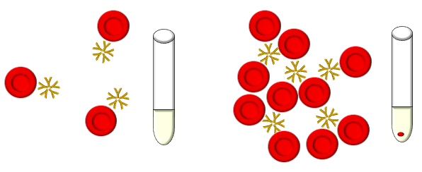 IgG however, only has a reach of 12-14 nm and in most instances is unable to overcome the distance between two red cells.
Figure 2: Since most of the antibodies we are interested in are IgG in nature, a specific reagent containing IgG antibodies with specificity against the Fc or constant region of other IgG molecules was developed. The binding of this anti-IgG antibody (also known as antihuman globulin or Coomb’s reagent) results in the formation of a bridge between red cells that have IgG bound to their surface. This in turn creates agglutinates that are visible to the human eye. Additional details on AHG or Coomb’s testing is provided in following section.
Figure 3: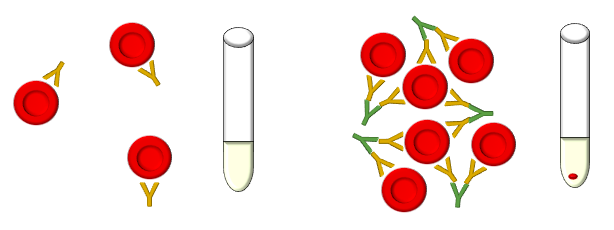
- Polymers
- Examples of polymers include albumin and polyethylene glycol (PEG). Polymers, or potentiators, can enhance agglutination through the reduction of the zeta potential. Both albumin and PEG are thought to act by displacing water molecules in between red cells, which in turn decreases the zeta potential and allows the red blood cells to come into closer contact.
- Proteolytic enzymes
- Examples of enzymes include ficin, papain, bromelin and trypsin. Enzymes can enhance agglutination by removing the negatively-charge sialic acid from the red cell membrane, which also reduces the zeta potential. Enzymes can also remove red cell glycoproteins reducing the steric hindrance on antibodies interacting with antigens.
- Enzymes typically enhance reactions involving antibodies targeted against the Rh and Kidd blood type antigens, while reducing or eliminating reactions involving antibodies against Duffy and MN antigens.
Table 1: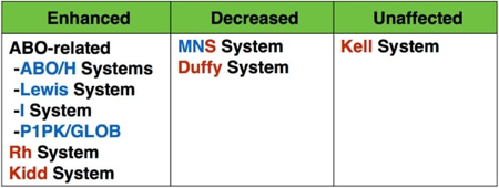
Next page: Serological endpoints: Agglutination - Additional Notes on Coombs Testing