
Antibody Panels: Getting Oriented:
The purpose of the antibody panel is to determine the specificity of any red blood cell antibody or antibodies that were identified initially on the antibody screen. This is accomplished by checking for reactivity between patient plasma and a larger number of reagent red blood cells which collectively express antigens which patients frequently make antibodies against.
Both antibody screens and antibody panels share a common structure: each row represents reagent red blood cells collected from a single individual (Figure 1). The term “Donor” on the left-hand side of the table in this Figure refers to the individual from whom these reagent cells were collected, not the individual whose blood will be used for transfusion.
The “+” and “0” signs denote whether or not the corresponding antigen, which is marked in the column header, is either expressed (+) or not expressed (0) by that cell. The combination of Rh antigens expressed by each cell is also summarized as a Wiener haplotype (e.g., R1R1, R1r, etc.) as described in the previous module.
The far-right column in Figure 1 labeled “Patient results” is where the reaction between the RBC and patient plasma is recorded. Reactions are graded from “0” to “4+” with “0” representing a negative reaction, and everything from “weak” to “4+” representing varying strengths of positive reaction. In the Figure below, strength of reaction by indirect antiglobulin testing is indicated, but other columns can be used for other types of testing if desired (e.g., after treatment of cells with proteolytic enzymes)
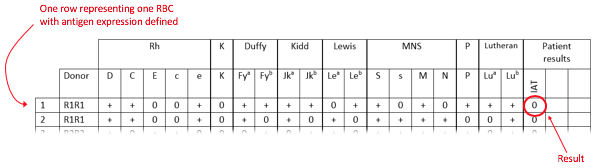
Figure 1: Example of a panel. One row represents one RBC with reactions against that RBC presented in the far-right column. For example, in row 1 (cell 1), the RBC is positive for D, C, and e antigens, but negative for E and c antigens. The patient’s plasma is non-reactive with this RBC as indicated by the “0” in the results column.
There are several considerations when interpreting a positive or negative reaction. Keep in mind that there are always exceptions to the rule and that only the most common interpretations will be presented here.
The reaction is negative. What does that mean?
If there is no reaction between the RBC and patient plasma, then the antibody which was first noted on the antibody screen is unlikely to be against any antigens that are known to be present on that specific RBC, especially antigens which are strongly expressed (otherwise we would have expected a positive reaction).
Because the lack of reactivity of patient plasma against a single cell is often sufficient to rule out specificity for multiple different antigens, performing exclusions with non-reacting cells is usually the first step when approaching a panel. When performing exclusions, it is very important to consider genetic zygosity.
The reaction is positive. What does that mean?
If the reaction between the RBC and patient plasma is positive, this suggests an antibody in the patient’s plasma is binding to an antigen expressed by that cell. The target of the antibody may include any antigen known to be present on the RBC (denoted with a “+” in that row) or may include uncommon antigens whose presence or absence are not indicated on the panel.
Remember: there are over 300 different RBC antigens in existence and it would be impossible to mark them all on a panel! Only those to which antibodies are most commonly detected are included.
The reaction of patient plasma against any one cell is not enough to identify which antigen it is reacting with; instead, a pattern must be established. This is described in more detail in the next section.
When is your antibody investigation complete?
While policies may vary between laboratories, a completed investigation is usually defined as one in which a) all positive reactions have been convincingly attributed to a specific antigen or collection of antigens, and b) antibodies to all other commonly, clinically significant antigens have been ruled out.
Common clinically significant antigens are usually defined as D, C, c, E and e (in the Rh system), K (in the Kell system), Jka and Jkb (in the Kidd system), Fya and Fyb (in the Duffy) and M, N and S (in the MNS system).
- Note: It is not necessary to exclude reactivity against every single antigen listed on an antibody panel. While antibodies against high-frequency antigens such as k or s are usually easy to exclude, it may not be possible to exclude antibodies against low-frequency antigens such as Lua, Kpa, Jsa, Cob, Wra, V or Cw. So long as all positive reactions are accounted for, and all other common clinically significant antibodies are excluded, however, these additional rule-outs are not required.
Next page: Zygosity